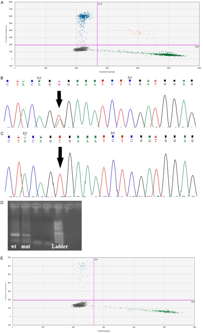
Figure 3.
These figures shows samples from one patient with malignant melanoma (primary biopsy: A; C versus distant metastases B; D; E). Sample (A) was positive for the BRAF V600E mutation only with droplet digital PCR. The levels of positive and negative droplets in sample (A) were 62.6 and 111 copies/μL respectively and these figures were used to calculate the concentration of target DNA. Sample (C): was negative for the BRAF V600E mutation using Sanger sequencing. Sample (B) was positive for the BRAF V600E mutation by Sanger sequencing (B), allele-specific PCR (D) and droplet digital PCR (E). The levels of positive and negative droplets in sample (D) were 6.21 and 36.6 copies/μL respectively. The blue cluster (FAM-fluorescence signal) represents droplets that were positive for the BRAF V600E mutation and the green cluster (HEX-fluorescence signal) represents droplets that were negative for BRAF V600E. The brown cluster represents double-positive droplets containing both wild-type and mutated DNA and the grey cluster represents droplets containing neither template.

