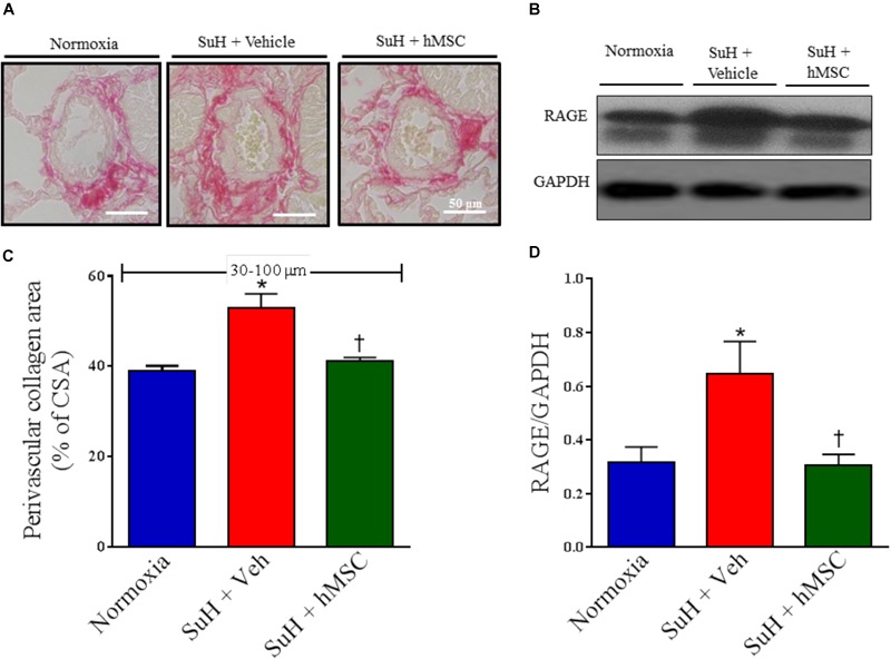
FIGURE 3.

Effects of the hMSC therapy in the pulmonary vascular extracellular matrix deposition. (A) Picrosirius red staining. (B) Shows the Western blot analyses of RAGE in lungs from all animal groups. GAPDH was used for normalization. (C) Perivascular collagen area, and (D) quantification of RAGE expression. Each column and bar represent the mean ± SEM (n = 5–7 mice per group). ∗P < 0.05 compared with normoxia group; †P < 0.05 compared with SuH group treated with vehicle. Ordinary one-way ANOVA with multiple comparisons. hMSC, human mesenchymal stem-cell; SuH, SU5416/hypoxia; RAGE, receptor for advanced glycation end products; CSA, cross section area.
