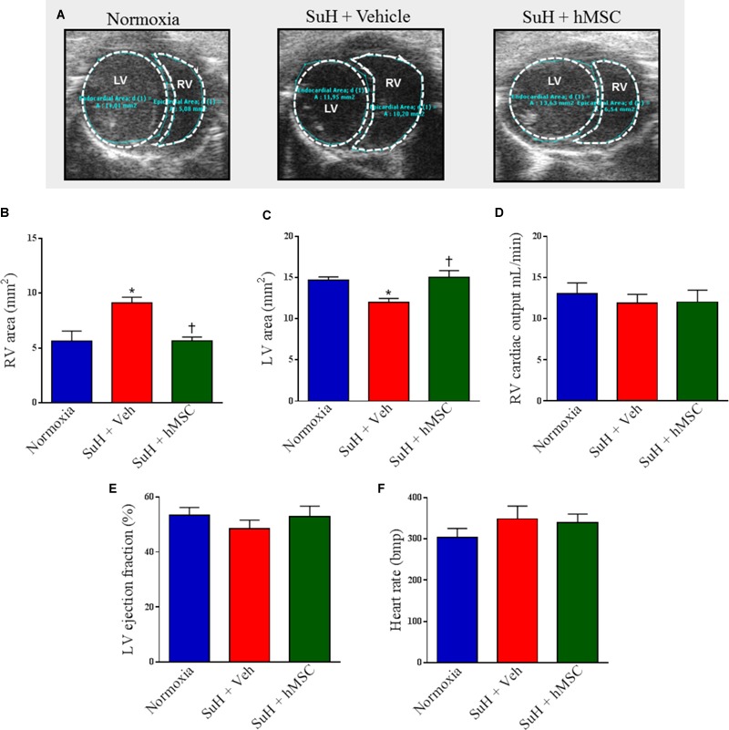
FIGURE 5.

Effects of the treatment with vehicle or hMSC on heart structure and function of SuH-PAH mice. (A) Representative images of parasternal short-axis views obtained by B-mode echocardiography (all end-diastolic), (B) right ventricle area, (C) left ventricle area, and (D) right ventricular cardiac output 28 days after protocol initiation. (E) Left ventricle ejection fraction and (F) heart rate. Each column and bar represent the mean ± SEM (n = 5–7 mice per group). ∗P < 0.05 compared with normoxia group; †P < 0.05 compared with SuH group treated with vehicle. Ordinary one-way ANOVA with multiple comparisons. hMSC, human mesenchymal stem-cell; SuH, SU5416/hypoxia; RV, right ventricle; LV, left ventricle.
