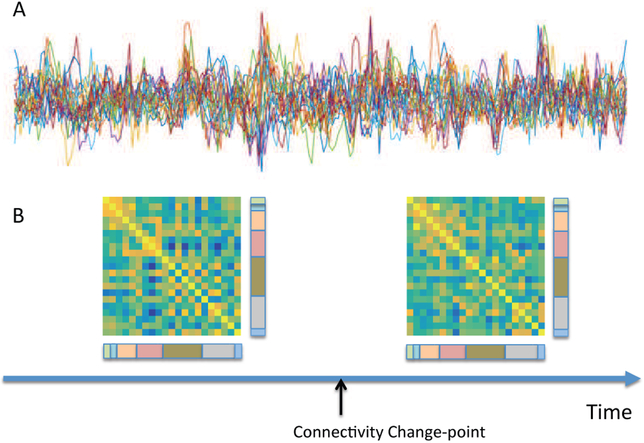
Figure 5.
Figure legend: (A) Resting-state fMRI time series from 21 regions of the brain from a single subject. (B) The results of a change point analysis ([109]) show a connectivity change point roughly half way through the scanning session. The two correlation matrices represent the brain state before and after the change point. Edges between visual components (color-coded as pink) and both cognitive control (olive green) and default mode (grey) components appear to be particularly variable. normalsize

