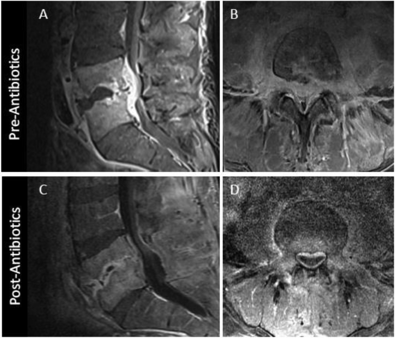
Figure 12.
MRI appearance of DOM before and 8 weeks after successful antibiotic treatment. Sagittal (A) and axial (B) T1 post contrast images coned in to the L4-L5 level show advanced DOM at this level with liquification of the L4-L5 disc, endplate erosion, and prominent ventral epidural and paraspinous spread of infection. Following 8 weeks of antibiotic therapy, axial T1 post contrast MR image (C) shows some progressive vertebral body erosion and height loss at L4-L5 with persistent abnormal marrow enhancement. Axial T1 postcontrast images (D) however, reveals significant decrease in epidural and paraspinous phlegmon, a more reliable indicator of interval response to therapy.

