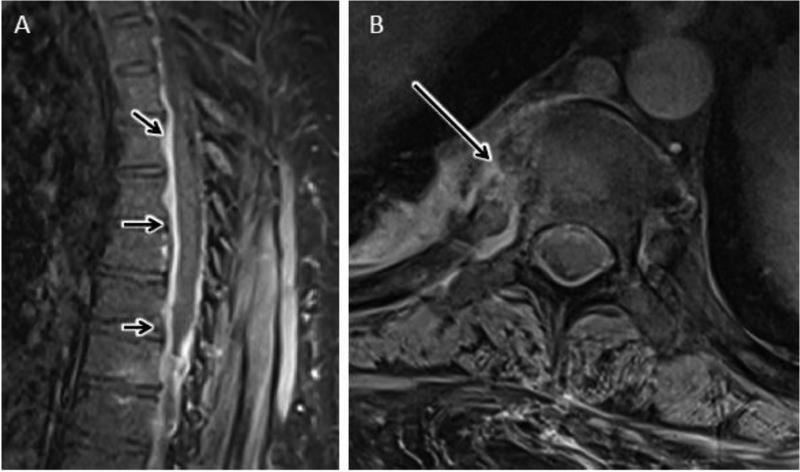
Figure 16.
Septic costo-vertebral joint in a 49 year-old male with elevated CRP and back pain. A) Sagittal T1 post contrast FS MR image of the thoracic spine shows longitudinally extensive ventral-predominant epidural thickening and hyperenhancement (short arrows) without evidence for DOM at any thoracic level. B) Axial T1 post contrast FS MR image at the T7 levels shows abnormal bone and peri-articular enhancement centered about the right 7th costovertebral joint, consistent with septic arthropathy.

