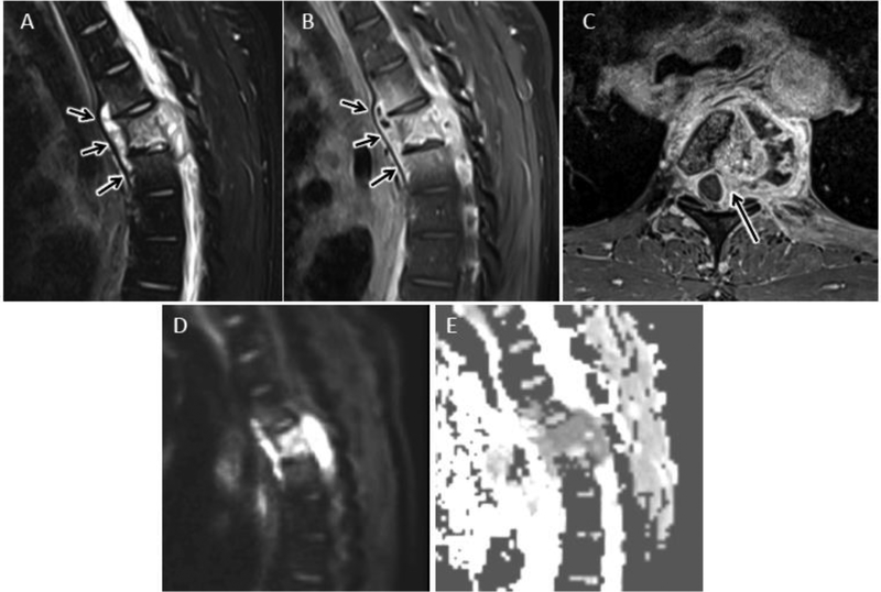
Figure 17.
Tuberculous spondylitis (TS) in a 31 year old male from central Africa presenting with several month history of cough, fever, night sweats, and 25 lb weight loss. Sagittal T2 FS (A) and T1 post FS (B) MR images reveal abnormal T2 hyperintensity and enhancement centered at T5 with prominent anterior subligamentous spread of infection (arrows). Note relative sparing of the T4–5 and T5–6 intervertebral discs. C) Axial T1 post contrast MR image shows T5 osteomyelitis with left more than right epidural extension (arrow) and irregular left paraspinous abscess. Sagittal DWI (D) and ADC map (E) reveals confluent reduced diffusion in regions of solidly enhancing infection. Percutanous biopsy confirmed the suspected diagnosis of TS.

