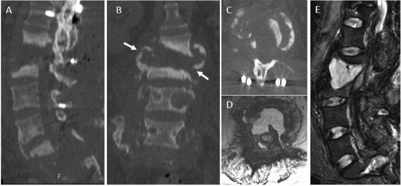
Figure 20.
Spinal neuroarthropathy (SNA) as a mimic of discitis-osteomyelitis in a 58-year-old male with multilevel destructive SNA 7 years after traumatic paraplegia, most pronounced at L2–L3 and S1-S2. CT scan with sagittal (A), coronal (B), and axial (C) plane images and MRI with axial (D) and sagittal (E) T2w images show complete destruction of the discovertebral unit at L2–3 with osseous debris extending beyond the vertebral body (arrows in B) as well as presence of an intervertebral collection. Extensive destruction of the sacrum is also seen.

