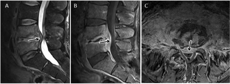
Figure 4.
Advanced lumbar DOM with extension beyond the discovertebral complex. Sagittal T2 (A), sagittal (B), and axial (C) T1 post contrast MR images coned in to the lower lumbar spine show characteristic MR features of L4-L5 DOM. In addition, there is prominent ventral epidural extension of phlegmonous infection (arrow in A-C) contributing to spinal canal stenosis. Axial T1-post contrast image (C) best demonstrates the extensive ventral and anterolateral paraspinous extension of infection without drainable fluid collection.

