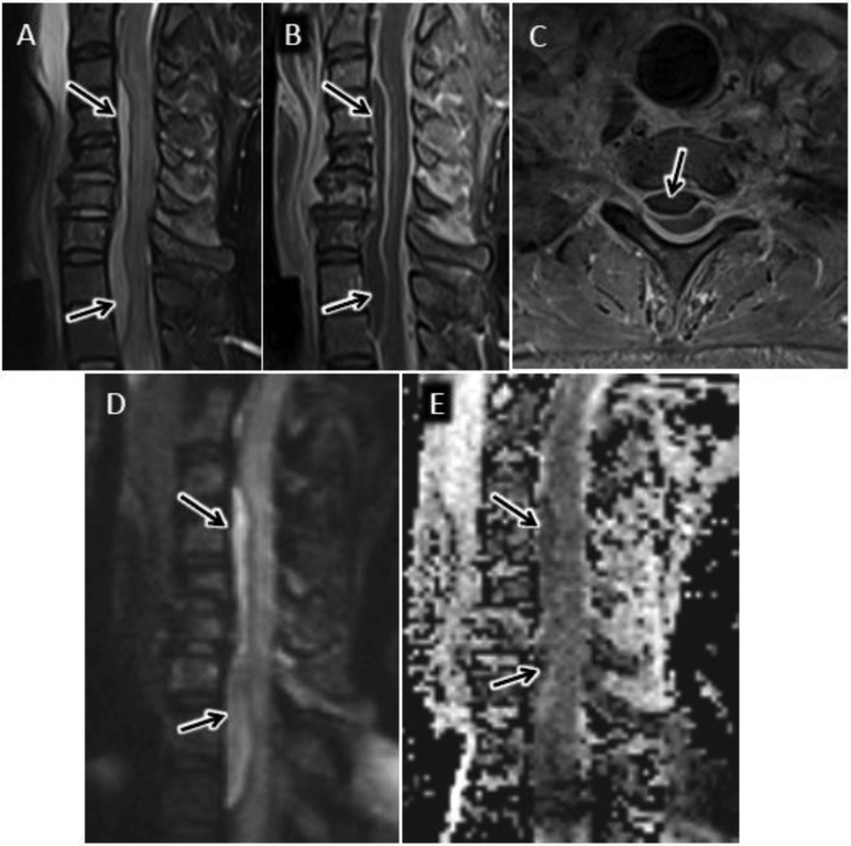
Figure 6.
Cervical epidural abscess with reduced diffusion. 48 year-old man with history significant for intravenous drug abuse presents with progressive weakness and quadriplegia. Sagittal T2-w (A) and sagittal (B) and axial (C) T1-w post contrast MR images of the cervical spine reveal a longitudinally extensive ventral epidural fluid collection with peripheral enhancement and spinal cord compression. Sagittal DWI (D) image and corresponding ADC map (E) through the same level demonstrate abnormal reduced diffusion associated with this epidural abscess.

