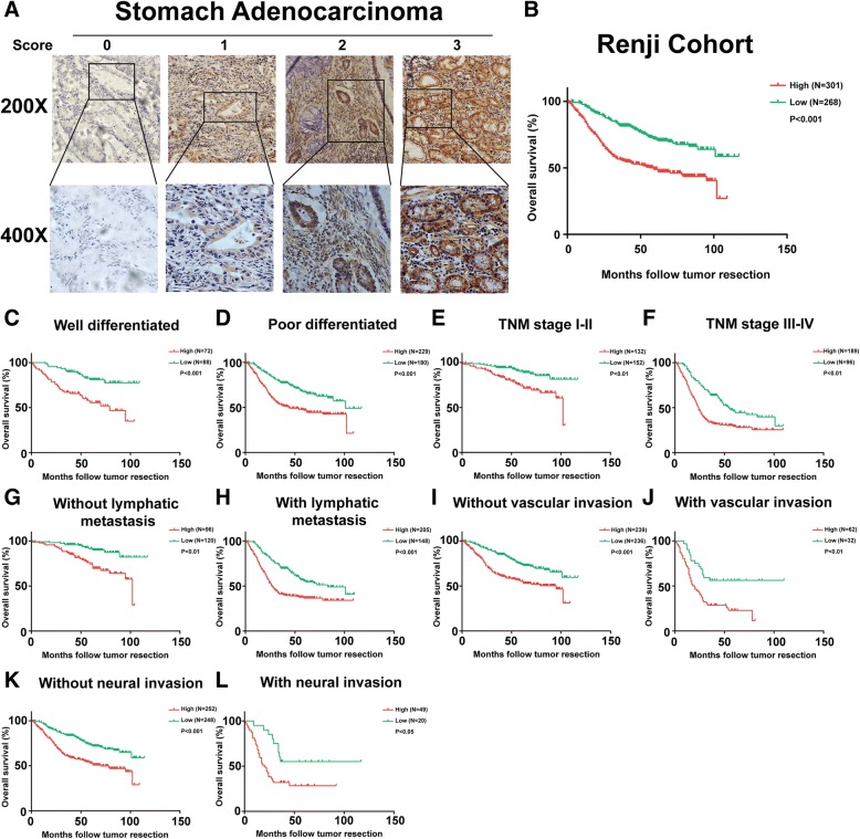
Fig. 2.
Association between GRINA expression and prognosis in gastric cancer. a GRINA protein expression was assessed immunohistochemically in TMAs containing 569 gastric cancer samples. GRINA was located in the plasma membrane and cytoplasm. Scoring was conducted according to the ratio of positively stained cells: 0–5% scored 0; 6–35% scored 1; 36–70% scored 2; and more than 70% scored 3, and staining intensity: no staining scored 0, weakly stained scored 1, moderately stained scored 2, and strongly stained scored 3. b The Kaplan-Meier analysis showed that the patients with higher GRINA expression had poorer overall survival. c-l The Kaplan-Meier curves of overall survival related to histological differentiation, TNM stage (stage I and II, stage III and IV), lymphatic metastasis, vascular invasion, and neural invasion according to the GRINA levels in 569 gastric cancer samples

