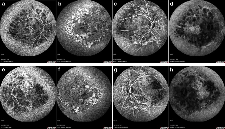
Fig. 4.
FFA and ICGA. In the early phase of FFA, the background fluorescence was observed around the disc decreased with choroidal vascular exposed, and diffuse pinpoint hyperfluorescent leakage was noticed in the peripheral parts reached to the equator (a: right eye; e: left eye). In the late phase of FFA, the hypofluorescence around the disc grew to hyperfluorescence formed to be “honeycomb” appearance in the posterior pole (b: right eye; f: left eye). In the early phase of ICGA, choroidal vascular exposed in the posterior pole around the disc (c: right eye; g: left eye). In the late phase of ICGA, massive hypofluorescence of choroidal was noticed in the posterior pole around the disc, while hyperfluorescence was observed in the peripheral parts reached to the equator (d:-right eye; h: left eye)

