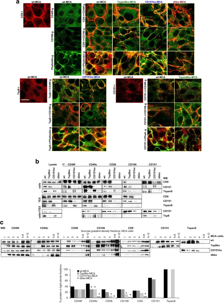
Fig. 4.
Adhesion molecule location and association with tetraspanins in wt- and ko-MCA tumor cells. a Colocalization of CD49f, CD106 and F-actin (Phalloidin) with CD9, Tspan8 and CD151 was evaluated by confocal microscopy. Representative examples of single staining and overlays are shown (scale bar: 10 μm); b coIP of CD49f, CD49c, CD56 and CD106 with CD9, CD151 and Tspan8 in wt- and ko-MCA cells, TEX and wt-TEX-treated cells; c WB of wt- and ko-MCA cell lysates after sucrose gradient centrifugation, where the light fractions (1–4) were tested individually and medium dense fractions 5–8 and dense fractions 9–12 were pooled; representative examples are shown and the mean intensity of light fractions (1–4) in comparison to all fractions (3 assays) is shown; significant differences in the recovery in light fractions between wt- and ko-MCA cells: *. CD49f most prominently colocalizes with CD9 and Tspan8 in CD151ko-MCA, whereas CD106 preferentially colocalizes with CD151. F-actin associates with CD9 and Tspan8, but hardly with CD151. CoIP confirmed preferential association of CD49f and CD49c with Tspan8 and of CD56 and CD106 with CD151 in cells and, less pronounced, TEX and wt-TEX-treated cells. In MCA cells, the association of adhesion molecules with tetraspanins is accompanied by pronounced recovery in light density (TEM) fractions

