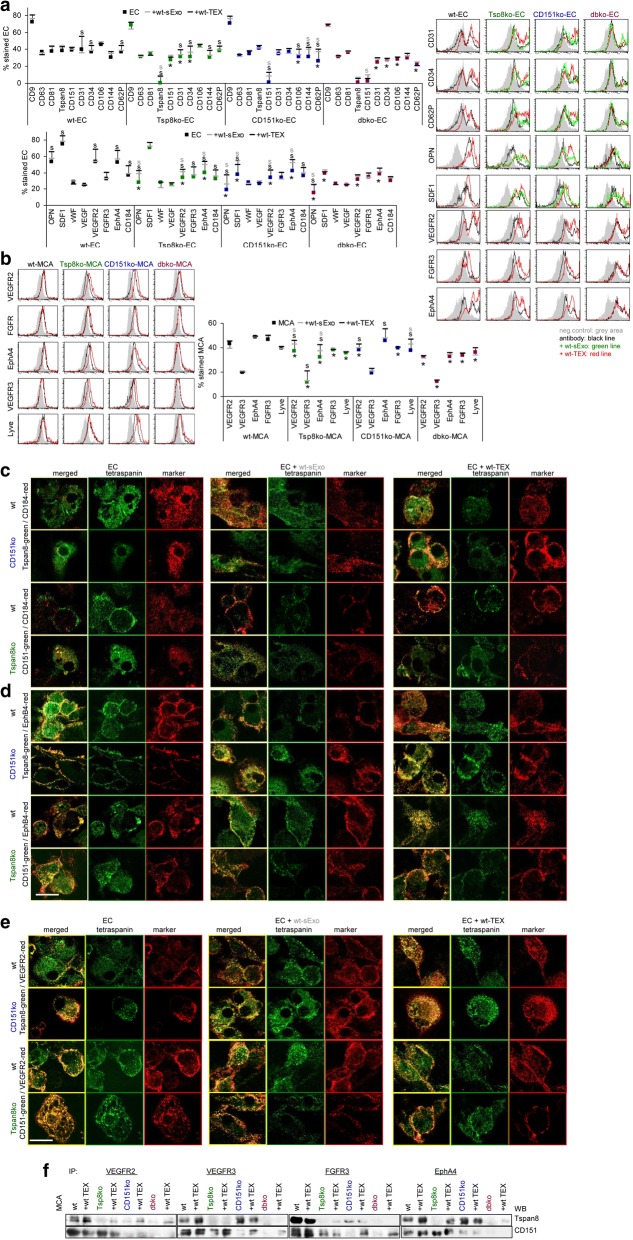
Fig. 8.
Tspan8, CD151 and angiogenesis in ko mice and ko MCA tumors. Dispersed lung cells were cultured for 3wk-4wk to enrich and expand EC. EC were cocultured with wt-sExo or wt- and ko-TEX for 48 h–72 h. a EC flow-cytometry analysis of tetraspanins, EC markers, chemokines and angiogenic receptors including representative examples and (b) of MCA angiogenic receptor; a,b mean % stained cells (3 assays), significant differences between wt- and ko-EC or -MCA cells: *, significant differences by coculture with sExo: s (grey) or TEX: s (black); c-e confocal microscopy of wt-, Tspan8ko-, and CD151ko-EC cultured in the absence or presence of wt-sExo or -TEX and stained with anti-Tspan8 or anti-CD151 and counterstained with (c) anti-CD184, d anti-EphB4, e anti-VEGFR2, single staining and overlays are shown (scale bar: 10 μm); f untreated and wt-TEX-treated wt- and ko-MCA lysates were precipitated with anti-VEGFR2, -VEGFR3, -FGFR3 and EphA4. Dissolved precipitates were blotted with anti-Tspan8 or anti-CD151. Representative WB examples are shown. Ko-EC express CD31, CD34, OPN, VEGFR2, and EphA4 at a reduced level; in CD151ko-EC additionally CD106 and CD62P expression is reduced. Wt-sExo promote OPN, VEGFR2 and EphA4 expression in ko-EC; wt-TEX activate EC marker, OPN, SDF1, VEGFR2, EphA4 and CD184 expression in wt- and ko-EC. In ko-MCA cells, wt-sExo and -TEX support VEGFR2, VEGFR3 and EphA4 expression. Upregulated expression, accompanied by colocalization and coimmunoprecipitation, might well account for Tspan8- and CD151-TEX promoted EC activation

