
Fig. 2.
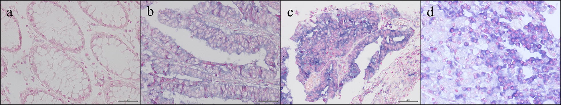
HOTAIR expression in colon tissue samples: a negative HOTAIR expression in colonic mucosa (×20); b positive HOTAIR cytoplasmic expression in adenomatous dysplastic area (×20); c positive HOTAIR cytoplasmic expression in CRC sample (×20); d detail of lymphocytes staining in tumor microenvironment of CRC sample (×60)