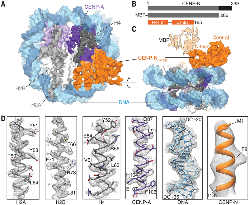
Fig. 1. Structure of the human CENP-N/CENP-A nucleosome complex.
(A) Cryo-EM density map of the hCENP-N1–286/CENP-A nucleosome complex viewed down theaxis of the DNA supercoil. (B) Schematicof the functional domains of CENP-N known to bind the CENP-A nucleosome (gray) and CENP-L (black) (top panel). The CENP-N construct used for the present structural analysis (hCENP-N1–286) and the regions of the sequence whose structure we report here [N-terminal domain: residues 1 to 81, and central domain: residues 101 to 185; hCENP-N(1–185)] are shown in the middle and bottom panels, respectively. (C) Cryo-EM density mapof the hCENP-N1–286/CENP-A nucleosome complex as viewed from the side, at an orientation 90° to the view shownin (A). This view also depicts the extra density connected to the N-terminal domain that we assign to MBP, shown with lighter shading. (D) Representative regions of the cryo-EM density mapto illustrate map quality (from left to right) for canonical histones H2A, H2B, and H4, centromere-specific H3 variant CENP-A, nucleosomal DNA, and CENP-N.

