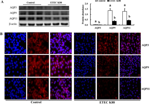
Figure 6.

Effect of enterotoxigenic Escherichia coli (ETEC) K88 challenge on the protein expression of intestinal aquaporins (AQP) in IPEC-J2 cells. Cells were challenged or not with ETEC K88 for 3 h. Then, treated cells were collected for western blotting analysis (A). Results are mean ± SEM (n = 6). a,bMeans with different superscripts between columns differ (P < 0.05). For immunofluorescence analysis (B), cells after treatment were fixed and incubated with primary antibodies (rabbit polyclonal antibodies to AQP3 [Bioss Antibodies, Beijing, P. R. China], AQP9 [AVIVA Systems Biology Corp, San Diego, CA], AQP11 [FabGennix International Inc, Frisco, TX]; red) followed by incubation with Alexa Fluor 568 goat anti-rabbit secondary antibody (Invitrogen, Merelbeke, Belgium). Nuclei (blue) were stained with 4',6-diamidino-2-phenylindole in ProLong Gold mounting medium (Invitrogen). Representative images (B) were taken with a Laser Confocal Microscope (Carl Zeiss AG, Heidenheim, Germany). Original magnification, 400x.
