Figure 2.
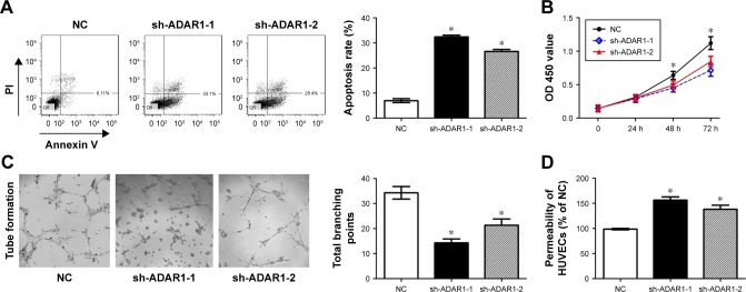
ADAR1 affected HUVEC cell apoptosis, proliferation tube branching formation and cell permeability.
Notes: (A) Cell apoptosis rates detected by flow cytometry. About 0.5–1×10⁶ cells were stimulated to induce cell death. (B) Cell proliferation was detected with a colorimetric assay using the CellTiter 96 AQueous One Solution. The absorbance value (OD) of each well was measured at 450 nm. (C) Image assay for tube formation analysis. HUVECs were plated on Matrigel in the presence of HUVEC media containing 5% bovine calf serum cells incubated with 25 ng/mL of hepatocyte growth factor to induce formation tubes in serum-reduced media. (D) Permeability assay of HUVECs. Values are expressed as mean ± SE. *P<0.05, as compared to the control.
Abbreviations: ADAR1, adenosine deaminase acting on RNA 1; HUVEC, human umbilical vein endothelial cell; NC, negative control; PI, propidium iodide.

