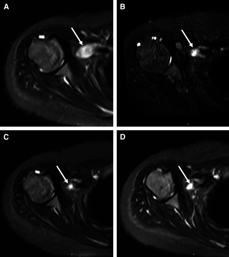
Fig 4.
Sequential axial fluid-sensitive magnetic resonance images at the level of the right glenohumeral joint performed (A) at diagnosis, and after (B) 8 weeks, (C) 16 weeks, and (D) 28 weeks of treatment illustrate the progressive decrease in size of the right distal brachial plexus lesion (arrow) over time.

