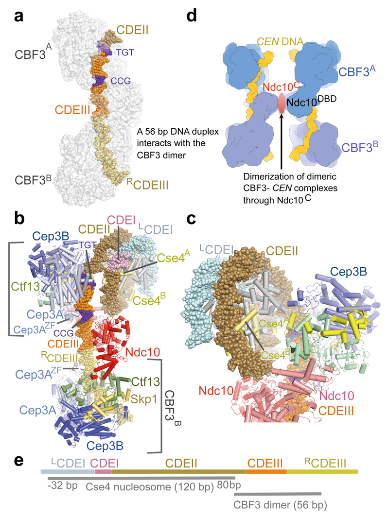
Figure 4. Model of CBF3−Cse4 nucleosome complex.
(a). Model of dimeric CBF3 with nuclease resistant 56 bp DNA segment. CBF3 is represented with a transparent molecular surface. (b) CBF3−Cse4−CEN structure modeled on the cryo-EM CBF3−CEN complex and the CENP-A crystal structure 40 (PDB 3AN2). The two CENP-A subunits (labeled as Cse4A and Cse4B) are highlighted. (c) Close up of the view of the CBF3−Cse4−CEN model showing close proximity of the N-terminus of Cse4A and Ctf13. (d) Cartoon showing possible pairing of CBF3−CEN complexes mediated by dimerization of Ndc10C domains. (e) Schematic of the proposed CBF3−Cse4 nucleosome complex at the CEN3 locus. CDEI, CDEII and CDEIII, and the DNA contact regions of Cse4 and CBF3 are drawn to scale. For Cse4, the start and end of the binding site are shown, -32 bp: 32 bp to the left of the LCDEI-CDEI junction, 80 bp: 80 bp to the right of the CDEI-CDEII junction.

