
Figure 1. act-4, tiam-1 and gex-3 are required for dendrite branching in PVD neurons.
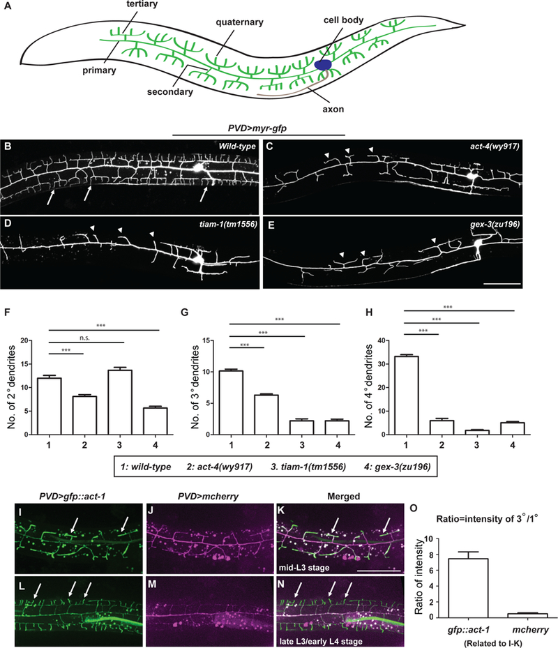
(A) Schematic showing the morphology of PVD dendritic arbors. Lateral view with anterior on left and dorsal up is shown. The same orientation is used for all fluorescent images in this work. (B-E) Representative confocal images showing the morphologies of PVD dendritic arbors labeled by a transgene ser2prom3>myr-gfp, in (B) wild-type, (C) act-4(wy917), (D) tiam-1(tm1556), and (E) gex-3(zu196) animals at late L4 stage. The most anterior and posterior parts are not shown. All PVD neuron fluorescence images in this work are labeled and presented by the same means unless otherwise noted. Arrows: 4o branches in the wild-type animals. Arrow heads: 2o branches that failed to form 4o or even 3o branches. Scale bar = 50 μm. (F-H) Quantifications of the number of 2o, 3o and 4o branches in a region 100 μm anterior to the PVD cell body. Data were obtained using 20 animals for each genotype and are presented as mean ± SEM. One-sided ANOVA with Dunnett’s test was used for statistical analysis. ***: p< 0.0001. n.s.: not significant. (I-N) Representative fluorescent images showing expression of GFP::actin during PVD dendrite branching and growth at indicated developmental stages. PVD was labeled by a transgene ser2prom3>mcherry. Arrows: dendrites with enriched GFP::actin. Scale bar = 50 μm. (O) Quantifications of the ratio of GFP::ACT-1 and mCherry intensity between 3o and 1o branches. Thirty-five 3o branches and 7 primary dendrites from 7 independent L3 stage animals were measured and the results are presented as mean ± SEM. See also Figure S1 and S2.