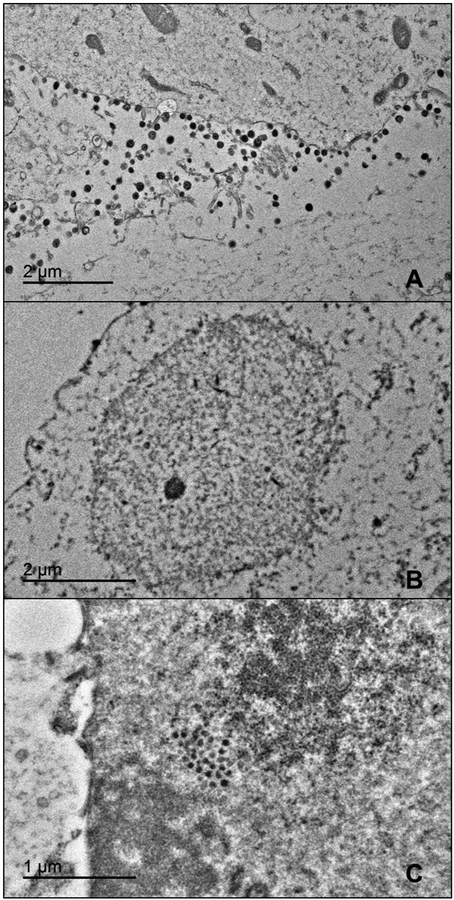
Fig. 2.
TEM of MDBK cells inoculated with BoHV-1 and treated with DMSO control (A), 50 μM αHT-106 (B), or 8 μM αHT-115 (C). Note the numerous extracellular virions in the control sample (A). No virions or capsids were observed in the αHT-106 treated (B) and αHT −115 treated samples (not pictured). Clusters of round electron dense intranuclear structures were very rarely present in the αHT-115 sample (C).

