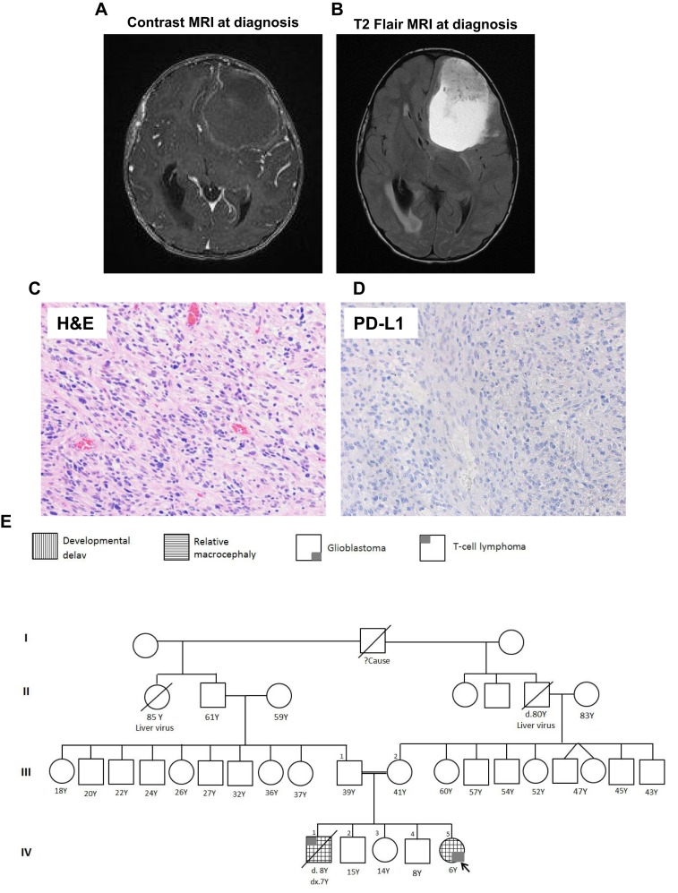
Figure 1.
Histological, radiological images of the patients with the family pedigree. MRI with (A) or without (B) contrast showing large intra‐axial left frontal predominately cystic complex mass with solid hemorrhagic components causing a severe mass effect is observed together with a right midline shift and obstructive hydrocephalus. (C): H&E staining demonstrating a densely cellular high‐grade glioma. (D): PD‐L1 immunohistochemistry with Dako 22C3 antibody is negative for immunoreactivity. (E): Four‐generation family pedigree of index patient. Abbreviations: H&E, hematoxylin and eosin; MRI, magnetic resonance imaging; PD‐L1, programmed death‐ligand 1.

