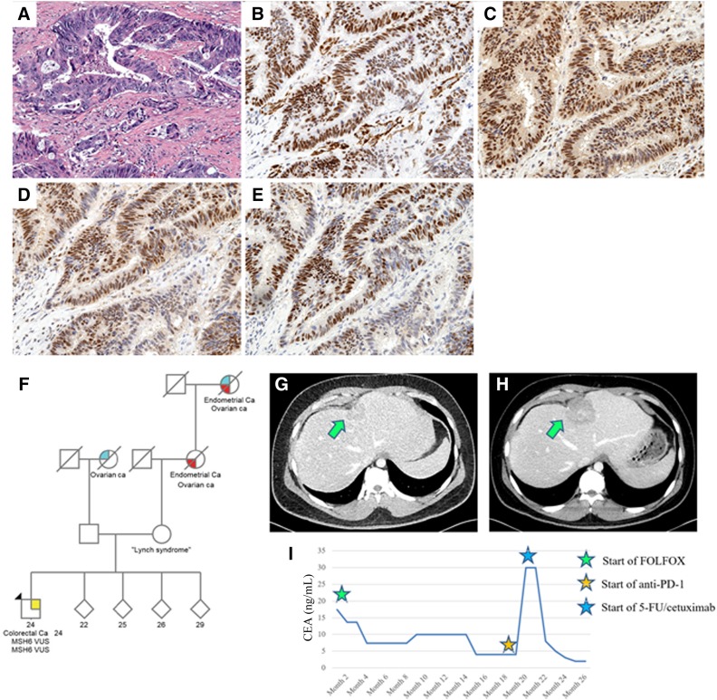
Figure 3.
Liver biopsy showing metastatic colorectal carcinoma (A). Mismatch repair protein staining showed intact MLH1 (B), intact MSH2 (C), intact MSH6 (D), and intact PMS2 (E) expression. (F): Family pedigree of patient 2. Proband is denoted by black arrow. Computed tomography of abdomen of patient 2 showing hepatic lesion prior to therapy (G) and after therapy (H). (I): CEA levels during treatment course with cytotoxic chemotherapy and with immunotherapy. Reference range for CEA is <5 ng/mL. Abbreviations: 5‐FU, 5‐fluorouracil; CEA, carcinoembryonic antigen; FOLFOX, folinic acid, 5‐fluorouracil, and oxaliplatin; PD‐1, programmed cell death protein 1; VUS, variants of uncertain significance.

