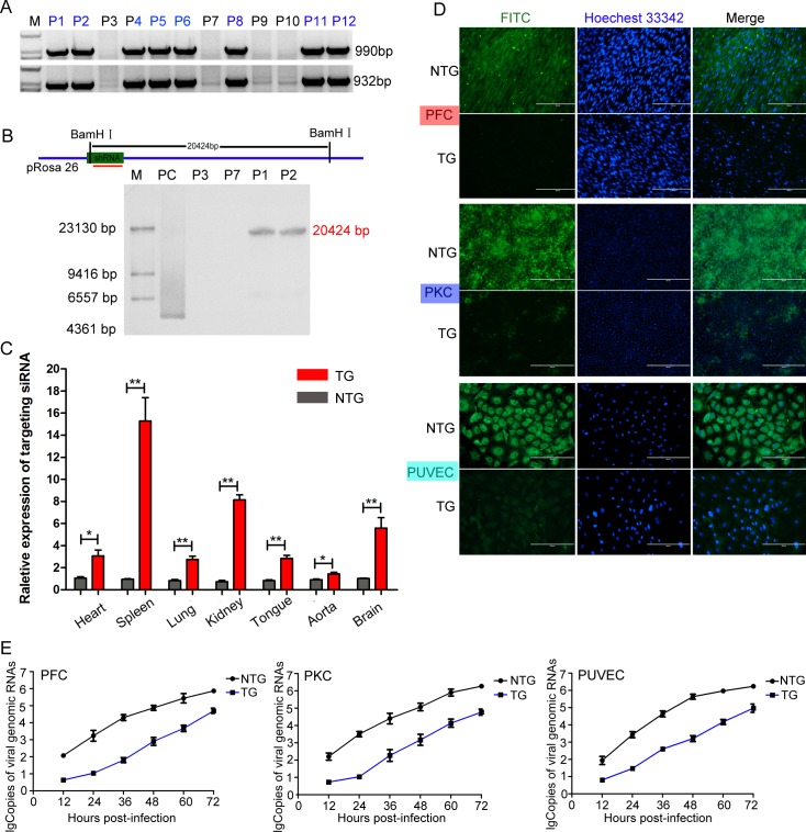
Fig 2. Generation of F0-generation TG pigs expressing the targeting siRNA.
(A) Genomic PCR analysis of the F0-generation to identify TG pigs. P1, P2, P4, P5, P6, P8, P11 and P12 indicate TG pigs, and P3, P7, P9 and P10 indicate NTG pigs. M: D2000. (B) Schematic for Southern blotting. The hybridization signals indicated that the shRNA gene was successfully integrated into the pRosa26 locus in the TG pigs. M: DNA maker. PC: positive control plasmid. P3 and P7 indicate NTG pigs, and P1 and P2 indicate TG pigs. (C) The relative expression levels of the targeting siRNA in various tissues and organs from TG pigs were determined by qPCR. The expression levels were analysed using the unpaired t-test (*p<0.05; **p<0.01). Error bars represent the SEM, n = 3. TG: transgenic pigs. NTG: wild-type pigs. (D) Virus resistance in three types of isolated primary cells from TG and NTG pigs was examined by IFA. The cells cultured in 24-well plates were inoculated with 200 TCID50 of CSFV (Shimen strain). At 72 hours post infection, the CSFV-infected cells were incubated with an E2-specific antibody (PAb) and then stained with fluorescein isothiocyanate (FITC)-labelled goat anti-pig IgG (1:100). Cells were analysed under fluorescence microscope. PFC: porcine fibroblast cells. PUVEC: porcine umbilical vein endothelial cells. PKC: primary kidney cells. (E) Genomic replication kinetics of CSFV in challenged TG and NTG cells at various time points post-infection. Error bars represent the SEMs, n = 3.

