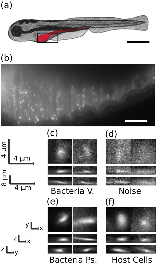
Fig 1. Images of bacteria in the intestine of larval zebrafish.
a) Schematic illustration of a larval zebrafish with the intestine highlighted in red. Scale bar: 0.5 mm. b) Single optical section from light sheet fluorescence microscopy of the anterior intestine of a larval zebrafish colonized by GFP expressing bacteria of the commensal Vibrio species ZWU0020. Scale bar: 50 microns. c) z, y and x projections from 28x28x8 pixel regions of representative individual Vibrio bacteria, d) non-bacterial noise, e) individual bacteria of the genus Pseudomonas, species ZWU0006, and f) autofluorescent zebrafish cells.

