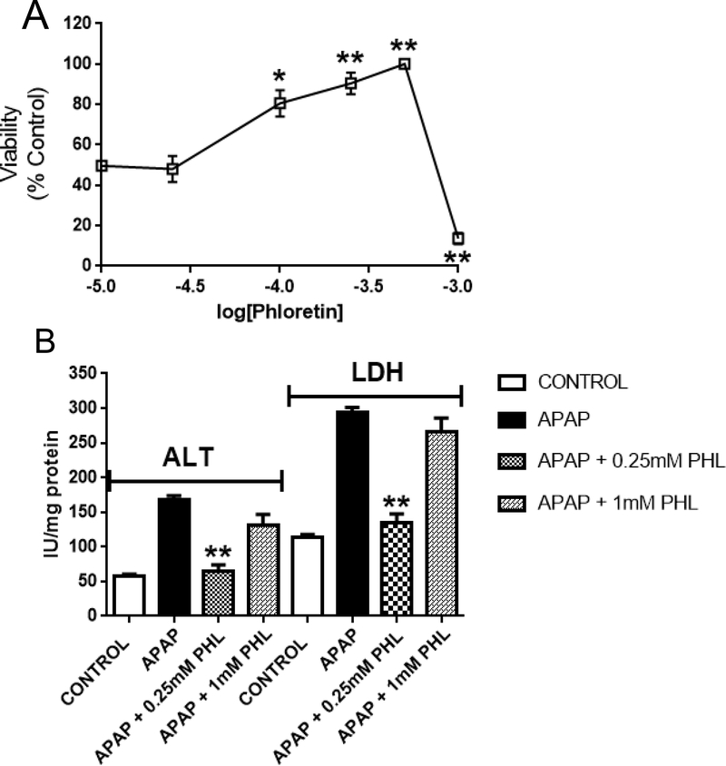
Figure 3.
(A) Effects of graded Phl concentrations (0.1 – 1.0 mM) on the viability of APAP (1.0 mM × 4 hrs)-exposed freshly isolated mouse hepatocytes. Data are expressed as mean percent of control ± SEM cell viability (n = 6–8). Exposure of hepatocytes to APAP alone (1.0 mM × 4 hrs) caused a decrease in mean cell viability of 48 ± 8% (see ordinate of Fig. 3A). Exposure of hepatocytes to phloretin concentrations ≤ 0.75 mM did not alter viability, whereas the 1.0 mM concentration caused a 59 ± 11% loss of viability (data not shown). (B) Concentration-dependent effects of phloretin (0.25 and 1.0 mM) on alanine aminotransferase (ALT) and lactate dehydrogenase (LDH) measured in incubation medium. Data are expressed as mean IU/mg protein ± SEM cell viability (n = 4–6). Statistically significant differences in treatment groups at *p<0.05 and **p<0.01 levels of significance.

