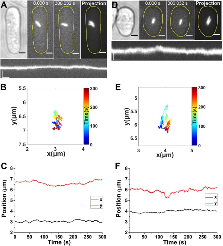
Figure 3.
Actin filaments and Myo52 are both indispensable to directed motion of cytoophidia. A) Time-lapse images of a cytoophidium in a LatA-treated cell. The cells were treated with 10 µM LatA for 30 min to disrupt the actin filaments before imaging. Far right, the maximum projection of the clip. Scale bars, 2 µm. Lower panel, kymograph of the cytoophidium. Scale bars, 2 µm (vertical) and 20 s (horizontal). B) Trajectory of the cytoophidium in the LatA-treated cell, with colored segments corresponding to the time points. C) Position of cytoophidium in the LatA-treated cell vs. time elapsed. D) Time-lapse images of a cytoophidium in a myo52Δ cell (in which the gene for Myo52 has been deleted). Far right, the maximum projection of the clip. Scale bars, 2 µm. Lower panel, kymograph of the cytoophidium. Scale bars, 2 µm (vertical) and 20 s (horizontal). E) Trajectory of the cytoophidium in the myo52Δ cell, with colored segments corresponding to the time points. F) x and y position vs. time of the cytoophidium in the myo52Δ cell.

