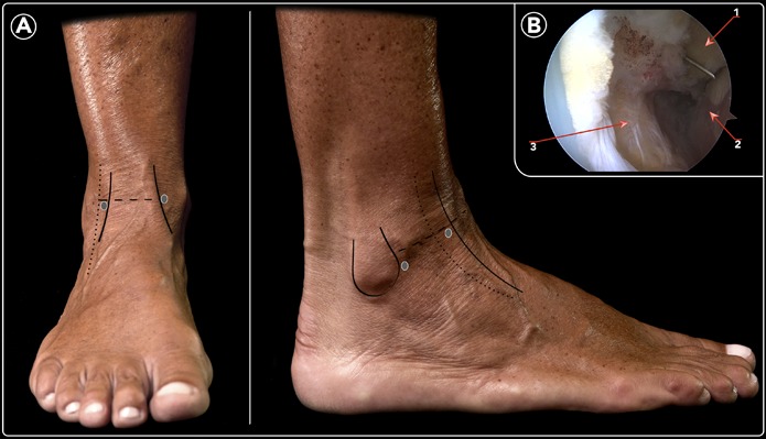
Fig. 1.

Anatomical landmarks and location of arthroscopic portals. Fig. 1-A Anterior and lateral views. See Video 1 for descriptions of landmarks. Fig. 1-B Arthroscopic view of the lateral gutter. A needle is introduced through the accessory anterolateral portal and observed at the lateral gutter. 1 = distal aspect of the lateral malleolus, 2 = lateral wall of the talus, and 3 = ATFL detached from the fibula.
