
Figure 3. CamKIIα‐transduced neurons colocalize with glutamate neurons.
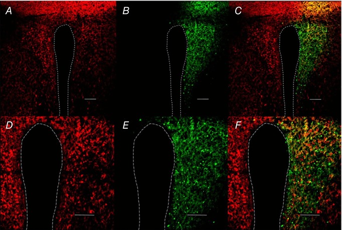
A, example of vGlut2‐Cre‐tdTomato‐positive neurons in a coronal cross‐section through the hypothalamus of a mouse (0.6 mm caudal to bregma). B, AAV2‐CamKIIα‐ChR2‐eYFP‐expressing PVN neurons. C, merge. A–C, scale bar = 100 μm. D–F, higher magnification (20×) of PVN CamKII‐transduced neurons; scale bar = 75 μm.