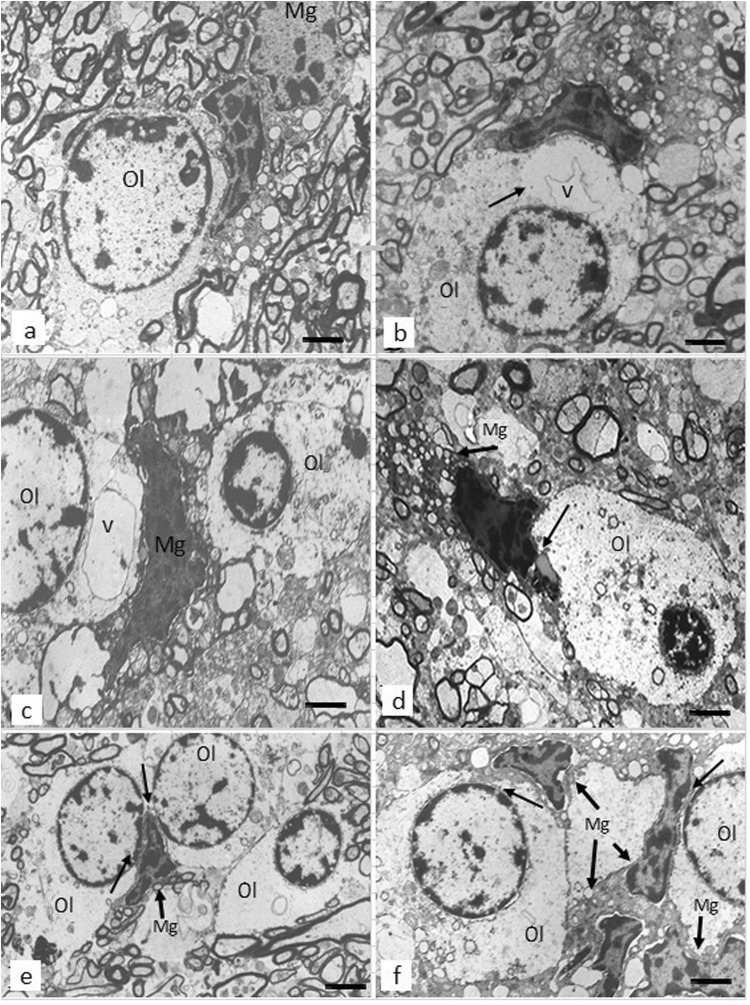
Fig. 1.
These micrographs from the prefrontal white matter show normal oligodendrocyte a and dystrophic changes of oligodendrocytes adjacent to microglial cells in SCZ brains b–f. Focal lysis of cytoplasm with the formation of vacuole in oligodendrocyte adjacent b to ameboid microglial cell and c to dystrophic microglial cell; d dystrophic microglial cell adjacent to lipofuscin granule in oligodendrocyte (arrow); e, f clusters of oligodendrocytes adjacent to microglial cell e and to microglial cell clusters f; e, f a contact of cytoplasm of microglial cells with the oligodendrocyte nucleus (arrows). Ol oligodendrocyte, Mg microglia, v vacuole (scale bar = 1 μm)

