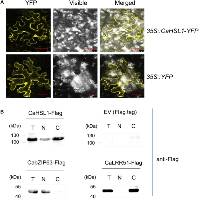
FIGURE 1.
The subcellular localization of CaHSL1. (A) CaHSL1 was localized to the plasma membrane and nucleus when transiently overexpressed in leaves of N. benthamiana that were infiltrated with GV3101 cells containing 35S::CaHSL1-YFP using 35S::YFP as control. The Agrobacterium-infiltrated N. benthamina leaves were harvested at 48 hpi, and counterstained by DAPI. Images were taken by confocal microscopy. Control (35S::YFP) showed signal throughout the cell. Bars = 50 μm. (B) The detection of CaHSL1 in the nucleus and cytoplasm by immunoblotting with total (T), nuclear (N), and cytoplasmic (C) proteins isolated from CaHSL1-FLAG or FLAG transiently overexpressing N. benthamina leaves using antibodies to FLAG. α-FLAG, FLAG antibodies.

