Figure 2.
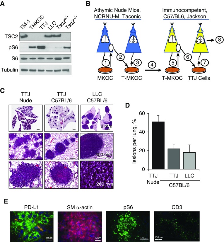
PD-L1 expression in Tsc2-null lung lesions in a metastatic immunocompetent mouse model of LAM. (A) Immunoblot analysis of Tsc2-expressing cells (TM1, Lewis lung carcinoma [LLC], and Tsc2+/+ mouse embryonic fibroblasts [MEFs]) and Tsc2-deficient cells (TMKOC, TTJ, and Tsc2−/− MEFs) with antibody against TSC2, pS6, S6, and tubulin. (B) Schematic representation of experimental procedures used to establish the Tsc2-null immunocompetent mouse LAM model. See text and the data supplement for details. (C) Representative H&E staining of lungs from nude mice injected with Tsc2-null TTJ cells, and C57BL/6 mice injected with either Tsc2-null TTJ or LLC cells. Animals were killed 18–21 days after injection, and lungs were inflated and stained with H&E. Scale bars: 2 mm, 200 mm, and 300 mm. (D) H&E images of the lungs were analyzed for the percentage of lesions per lung using Student’s t test for comparisons (n = 5 per each group, mean value ± SE), with P = 0.04 for TTJ nude versus TTJ C57BL/6, and P = 0.03 for TTJ nude versus LLC C57BL/6. (E) IHC analysis of pS6, SM α-actin, PD-L1 expression, and CD3+ T cells in C57BL/6 mouse lungs with Tsc2-null lung lesions (n = 5). Scale bars: 10 μm and 100 μm.

