Figure 3.
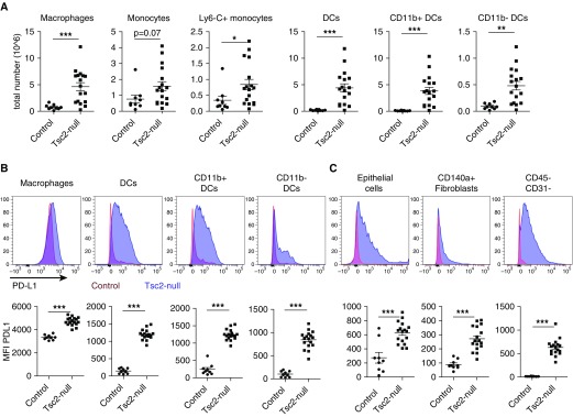
Antigen-presenting cells infiltrate lungs with Tsc2-null lesions and highly express PD-L1. (A) Lungs with Tsc2-null lesions taken from C57BL/6 mice killed ∼3 weeks after injection had increased numbers of macrophages, dendritic cells (CD11b+ and CD11b−), and monocytes (activated Ly6C+ and nonactivated Ly6C−). (B) Representative histograms show a shift in PD-L1 expression in lungs with Tsc2-null lesions (blue) compared with control lungs (red) in antigen-presenting cells, and quantification of PD-L1 expression via median fluorescence intensity (MFI) in these cell populations. (C) PD-L1 expression in CD45− stromal cell populations, including CD140a+ fibroblasts, epithelial cells, and CD31− cells (including Tsc2-null TTJ cells) in the mouse model of LAM. Statistical analysis was performed using Student’s t test with significance at *P < 0.05 (**P < 0.01, ***P < 0.001, n ≥ 5 mice, n = 2 experiments). DC = dendritic cell.

