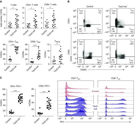
Figure 4.
T cells infiltrate lungs with Tsc2-null lesions and highly express PD-1. (A) Lungs with Tsc2-null lesions taken from C57BL/6 mice killed ∼3 weeks after injection have increased CD4+ and CD8+ T cells, CD4+ and CD8+ effector/effector memory T (TEM) cells, and regulatory T cells (TREGS), presented as a percentage of total CD4+ and CD8+ T cells. (B) Representative flow-cytometry diagrams illustrate the increase in CD4+ and CD8+ TEM cells in lungs with Tsc2-null lesions compared with controls. (C) Quantification of overall PD-1+ CD4+ and CD8+ T cells, and representative plots showing PD-1 expression on TEM cells in control lungs (red) compared with lungs with Tsc2-null lesions (blue). Statistical analysis was performed using Student’s t test with significance at *P < 0.05 (***P < 0.001, n > 5 mice, n = 2 experiments).

