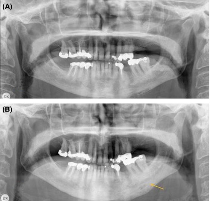
Figure 2.

Radiological examinations of a 71‐year‐old woman with diffuse sclerosing osteomyelitis of the left side of the mandible for two years, treated with corticosteroids and clindamycin for two years. A, Orthopantomogram before treatment with denosumab revealing sclerosis, resorption, and periosteal apposition of the left side of the mandible. B, Orthopantomogram taken after 12 mo with denosumab treatment showing less radiolucency and maturation of the bone in the area of periosteal apposition
