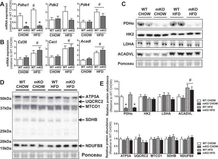
Fig. 4.
Metabolic and mitochondrial proteins in HFD-fed PDHmKO and WT mice. WT and PDHmKO mice were fed either a chow or a HFD for 12 wk. Transcript abundance of Pdha1, Pdk2, and Pdk4 (A) and Cd36, Cact (Slc25a20), and Acadl (B) in skeletal muscle from chow- or HFD-fed PDHmKO and WT mice, normalized to Ppib (Chow, n = 5/6; HFD, n = 6/7). Representative blot of PDHα, HK2, LDHA, ACADVL (C), and ATP5A, UQCRC2, MTCO1, SDHB, and NDUFB8 (D) protein abundance in skeletal muscle from chow- or HFD-fed PDHmKO and WT mice. Bar graphs show quantification of protein abundance in skeletal muscle relative to ponceau (Chow, n = 6/6; HFD, n = 6/6) (E). Data reported as means ± SE two-way ANOVA, #P < 0.05, main effect of diet, *P < 0.05, main effect of genotype (A–E). HFD, high-fat diet; PDHmKO, tamoxifen-inducible Pdha1 knockout mice; WT, wild-type.

