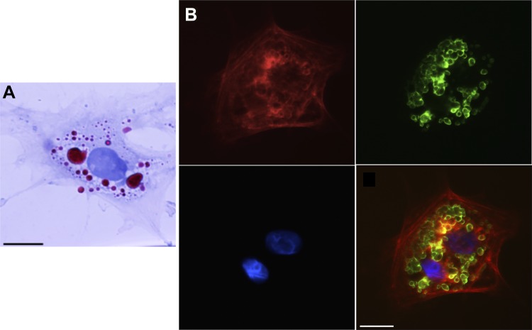
Fig. 4.
Oil red o (ORO) underestimates lipid droplet (LD) number and size within in vivo activated hepatic stellate cells (HSCs). A: HSCs isolated from day 14 postoperative rats that had undergone bile duct ligation were cultured for 12 h, and LDs were stained with ORO. B: cells from the same isolation were labeled with adipocyte differentiation-related protein (ADRP) primary antibody followed by fluorescein isothiocyanate-labeled secondary antibody (green) with nuclear stain 4',6-diamidino-2-phenylindole shown in blue. Smooth muscle α-actin labeling (red) was performed to verify cellular activation. Single channels and a merge image are shown. Representative images from >10 cells and >3 different cell preparations are shown. Scale bar = 10 μm. A minimum of 10 cells per time point were analyzed.

