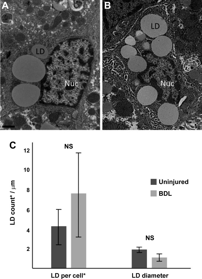
Fig. 7.
Hepatic stellate cells (HSCs) from bile duct ligation (BDL)-induced liver injury retain lipid droplets (LDs). BDL was performed as in materials and methods. After 10 days, livers were harvested and fixed as in materials and methods. Photomicrographs of HSC LDs from sham (A) and BDL-injured liver (B) were taken using standard transmission electron microscopy. Images shown are representative of >20 others. LDs from a single 100 nm plane (*) were counted, and diameters were measured using ImageJ software (C). A minimum of 10 cells per experimental condition were analyzed. n = 3 independent rats. Scale bar = 1 μm. NS, not significant.

