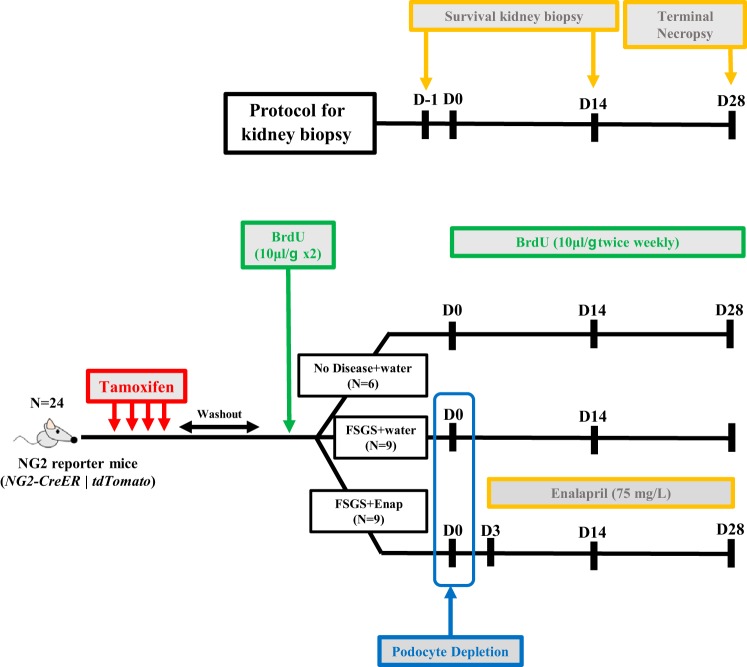
Fig. 1.
Schema of experimental design. Eight-week-old NG2-CreER tdTomato mice were given tamoxifen to label neural/glial antigen 2 (NG2) lineage cells with tdTomato, a red fluorescence protein (RFP) and then given a 4-wk tamoxifen washout period before initiation of experiments. Each mouse underwent two survival kidney biopsies in addition to the terminal necropsy of the kidney. There was a total of 24 animals randomized into 3 groups: focal segmental glomerulosclerosis (FSGS) animals given water (n = 9), FSGS animals given enalapril (Enap) (n = 9), and healthy mice that received a biopsy only (n = 6). Following the tamoxifen washout period, all mice underwent a survival baseline biopsy of the left kidney. This kidney tissue served as the baseline for each mouse. Following a 2-wk recovery period, 18 mice were randomly selected for induction of experimental FSGS (D0). This group of mice was further randomized on FSGS D3 to receive either drinking water or enalapril at 75 mg/l in drinking water. On D14 of disease, all 24 mice, including those without disease, underwent a second survival biopsy of the right kidney, which served as the tissue for D14 analysis. At D28, a terminal necropsy was performed. BrdU was administered throughout to identify cell proliferation. All animals received BrdU at 10 μl/g body wt before induction of FSGS and twice weekly after D0. D, day.

