Fig. 3.
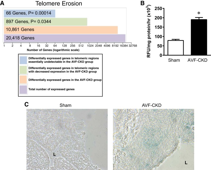
Telomere erosion and senescence-associated β-galactosidase (SA-β-Gal) activity/staining in the vein in the arteriovenous fistula (AVF)-chronic kidney disease (CKD) model. A: telomere erosion in the AVF-CKD vein, as shown by a disproportionate loss of gene expression in the telomeric regions, defined as 10 megabases from the start or end of each autosomal chromosome. Differentially expressed genes were defined by a false discovery rate of ≤0.05 when the AVF-CKD group was compared with the sham group. A total of 897 genes in the telomeric regions have decreased expression with a log2 fold change of <0; of these 897 genes, 66 have a log2 fold change less than −4. B: SA-β-Gal activity in sham veins and the vein of the AVF-CKD model at 1 wk. RFU, relative fluorescence units. Values are means ± SE; n = 5 in each group. *P < 0.0001. C: SA-β-Gal activity in frozen sections from a sham vein and the vein of the AVF-CKD model at 1 wk. Blue stain indicates cells with increased SA-β-Gal activity. The sham vein shows no staining, whereas the AVF-CKD model shows blue staining in the endothelium and media. L, lumen.

