
Fig. 4.
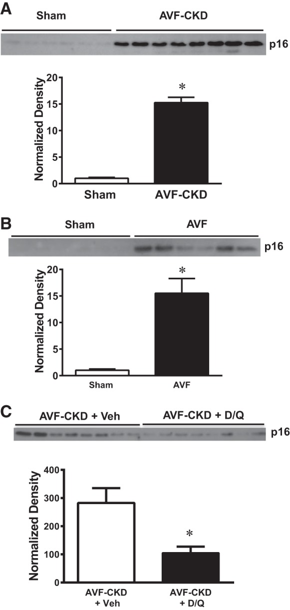
p16Ink4a expression in arteries in the arteriovenous fistula (AVF)-chronic kidney disease (CKD) model, in AVF without CKD, and in the vein in AVF-CKD following administration of senolytics. A–C: p16Ink4a protein expression at 1 wk in sham arteries and the artery of the AVF-CKD model (A), in sham veins and the vein of the AVF in mice with normal kidneys (B), and in the vein of the AVF-CKD model treated with vehicle (Veh) or dasatinib and quercetin (D/Q; C). Normalized expression is shown below each Western blot. Values are means ± SE; n = 6 sham and 8 AVF-CKD (A), 6 sham and 6 AVF-CKD (B), and 8 AVF-CKD + Veh and 8 AVF-CKD + D/Q (C). *P < 0.001 for A and B; *P < 0.05 for C.