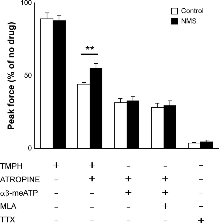
Fig. 4.
Contractility of bladder strips in presence of inhibitors. Peak contractile force in response to electric field stimulation (EFS) with TMPH (nAChR inhibitor), atropine (mAChR inhibitor), α,β-meATP (P2X1/3 desensitizer), MLA (α7 nAChR inhibitor), and TTX (Na+ channel blocker). The force was normalized with tissue weight and expressed as relative to the response to EFS in absence of inhibitors (N = 5–7 per group). Open and closed bars represent control and NMS groups, respectively. Means ± SE, **P < 0.01. α,β-meATP, α,β-methylene ATP; AChR, acetylcholine receptor; mAChR, muscarinic AChR; MLA, methyllycaconitine citrate; nAChR, nicotinic AChR; NMS, neonatal maternal separation; TMPH, 2,2,6,6-tetramethylpiperidin-4-yl heptanoate; TTX, tetrodotoxin.

