Fig. 5.
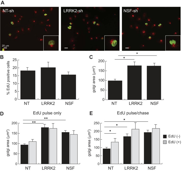
Golgi fragmentation in LRRK2 or NSF-deficient HK2 cells occurs independent of proliferation. A: epifluorescent images of EdU-labeled stable HK2 cell lines. Positivity for EdU integration into the cellular genome (green nuclei) is indicative of S-phase entry. Cells were costained with antibodies for endogenous gm130 (red) to mark the Golgi network in each cell. B: quantification of EdU-positive cells in each stable cell line. C: quantification of gm130-positive Golgi apparatus area in stable HK2 cell lines. D: quantification of gm-130-positive Golgi apparatus area in stable HK2 cell lines after a single 2 h pulse of EdU followed by immediate fixation and staining. E: quantification of gm-130-positive Golgi apparatus area in stable HK2 cell lines after a single 2-h pulse of EdU followed by 6-h medium chase before fixation and staining. The quantified data account for whether cells are positive (gray) or negative (black) for EdU labeling in both panels D and E. Error bars indicate standard deviation of values for triplicate experiments in which a minimum of 200 cells were quantified (*P < 0.05, **P < 0.005). EdU, 5-ethynyl-deoxyuridine; LRRK2, leucine-rich repeat kinase 2; HK2, normal human kidney cells; NSF, N-ethylmaleimide-sensitive fusion protein.

