-
A
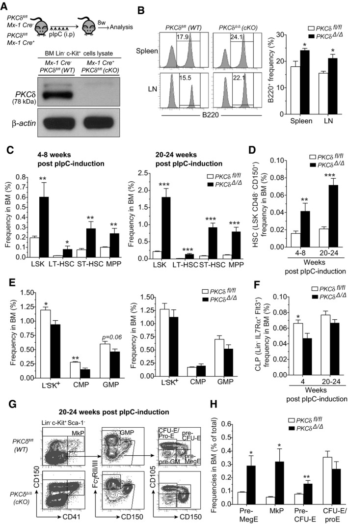
Experimental design. Representative Western blot analysis for detecting PKCδ protein in Lin−Kit+ BM cells from indicated mice at 8‐week post‐pIpC treatment shows absence of PKCδ protein in cKO cells.
-
B
FACS histograms show the frequency of B220+ cells in spleen and lymph nodes of cKO mice at 24‐week post‐pIpC treatment (n = 6–8 mice per genotype).
-
C
Increased frequency of HSPCs in the BM of control and cKO mice at 4–8 or 20–24 weeks after pIpC treatment (n = 8–9 mice per genotype and time point).
-
D, E
Frequency of HSCs (HSC‐SLAM) (D), and myeloid progenitor subsets (E) in the BM of control and cKO mice at 4–8 and 20–24 weeks after pIpC treatment (n = 8–9 mice per genotype and time point).
-
F
Frequency of common lymphoid progenitors (CLPs) in the BM of control and cKO mice at 4–8 and 20–24 weeks after pIpC treatment (n = 6 mice per genotype and time point).
-
G
Representative FACS plots show the gating strategy of MkP, Pre‐MegE, MkP, Pre‐CFU‐E, and CFU‐E/Pro‐E subpopulations in the BM of WT and cKO mice at 24 weeks after pIpC treatment.
-
H
Frequencies of indicated subsets in the total BM (n = 6 mice per genotype).
Data information: All data are presented as mean ± SEM, *
P < 0.05, ***
P < 0.01, and ***
P < 0.001, by repeated measures two‐way ANOVA analysis with Sidak's multiple comparison tests (B, D, and F) or by two‐tailed Student's unpaired
t‐test analysis (C, E, and H).

