-
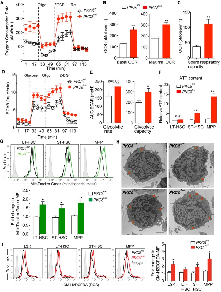
A
MitoStress test revealed increased oxygen consumption rates (OCR) in PKCδ‐deleted LSKs. BM LSK cells were sorted from WT and PKCδ cKO mice 12 weeks after pIpC treatment. OCR was measured at basal level and after sequential loading of ATP synthase inhibitor Oligomycin (350 nm), mitochondrial uncoupler, FCCP (10 μM), and electron transport chain inhibitor, Rotenone (1 μM) using Seahorse XF24 extracellular flux analyzer. Data are from three independent experiments (n = 9 mice). In each experiment, bone marrow cells from identical genotypes (n = 3 mice per genotype) were pooled and used for LSK cell sorting.
-
B, C
(B) Basal OCR was measured before inhibitors treatment (left); and the Maximal OCR capacity after FCCP treatment (right) (C). The spare respiratory capacity (SRC) of LSKs was calculated from the data shown in panel A (n = 9 per genotype, from three independent experiments).
-
D, E
(D) Glycolysis stress test. Extracellular acidification rates (ECAR) of LSKs were measured at basal conditions (unbuffered assay medium without glucose) and after sequential loading of glucose (7.5 mM), Oligomycin (350 nm), and 2‐deoxyglucose (50 mM; 2DG, a glucose analog) (E). AUC of baseline ECAR levels (left), and glycolytic capacity (maximal ECAR) was calculated from the data shown in panel (D) (n = 7–8 per genotype, from three independent experiments).
-
F
Relative ATP content in indicated HSPC subsets determined using an ATP assay kit (n = 6 mice per genotype).
-
G
Representative FACS histograms of MitoTracker Green fluorescence intensity on indicated HSPC subsets from control and cKO mice. Relative MitoTracker Green mean fluorescence intensity (MFI) is quantified below for n = 6 mice of each genotype.
-
H
Representative electron microscopy images (4,800× magnification) of control and PKCδ cKO HSPCs. Arrows indicate the mitochondria‐enriched regions (n = 3 mice per genotype). Scale bar represents 500 nm.
-
I
ROS levels in control and PKCδ cKO HSPCs as measured by FACS‐based CM‐H2DCFDA staining. Bar graph at right shows the relative CM‐H2DCFDA MFI. Data compiled from three independent experiments (n = 6–8 per genotype).
Data information: The statistical significance of difference was assessed using two‐tailed Student's unpaired
‐test analysis. All data are presented as mean ± SEM, *
< 0.001.