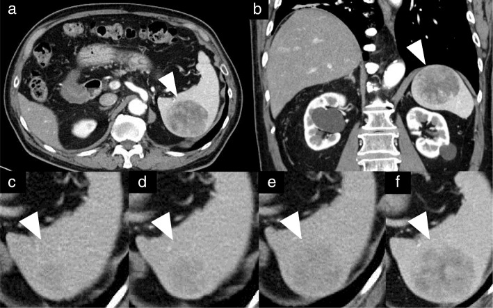
Fig. 1.
a and b: Computed tomography imaging shows a sharply circumscribed 50 mm tumor with slightly decreased uptake and a heterogeneous appearance in the spleen. c-f: A nodule appeared five years after thymectomy and enlarged gradually for three years thereafter. d: Six years after thymectomy. e: Seven years after thymectomy. f: Eight years after thymectomy. Arrows in all panels point to the metastatic lesion in the spleen

