Fig. 4.
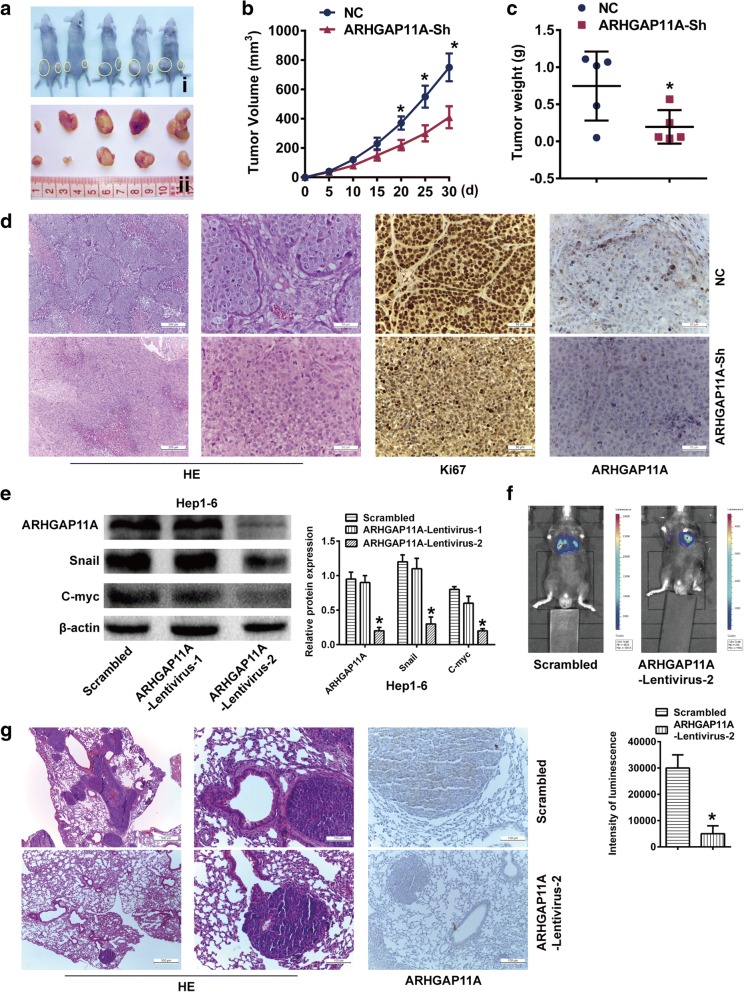
ARHGAP11A accelerates HCC tumor growth and metastasis in vivo. a ARHGAP11A-Sh-transfected MHCC97-H cells were injected subcutaneously into nude mice (n = 5). After 30 days, all mice were euthanized, and tumors were excised. Representative tumor photos are shown. i, NC left, and ARHGAP11A-Sh right. ii, NC up and ARHGAP11A-Sh down. b Growth curve of tumor volumes. *, P < 0.05 versus NC. c Tumor weight. *, P < 0.05 versus NC. d Representative images of tumor HE staining, and Ki-67 and ARHGAP11A immunohistochemistry staining are shown. Scale bars, 200 μm and 50 μm, respectively. e ARHGAP11A expression in Hep1–6 cells transduced with ARHGAP11A-lentivirus or a scrambled construct was analyzed by western blotting. f Representative bioluminescence images of C57BL mice 5 weeks after injection with cells transduced with ARHGAP11A-lentivirus or a scrambled construct are shown (n = 7). The luminescence intensity of lung metastases from luc-ARHGAP11A-Lentivirus-2- or scramble-transfected Hep1–6 cells was different. *, P < 0.05 versus the scrambled construct. g Lung metastatic nodules and ARHGAP11A expression in metastatic nodules were investigated with HE and immunohistochemical staining. Representative images are shown. Scale bars, 500 μm and 100 μm, respectively

