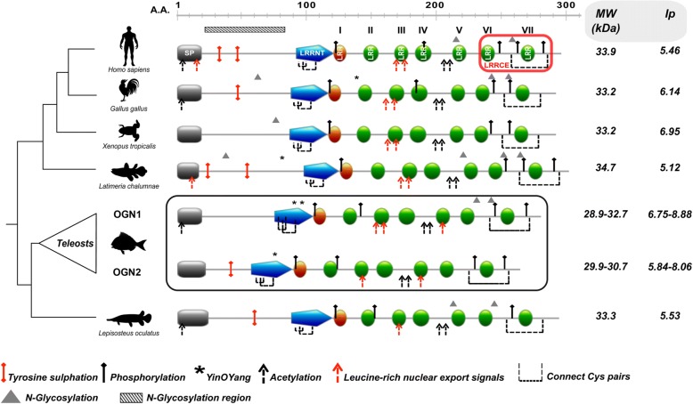
Fig. 1.
Dendrogram comparing OGN structural features from representative organisms of the main vertebrate lineages. Structural domains/motifs present in the amino acid sequences of OGNs from representative organisms are indicated as coloured blocks using the human OGN sequence as the reference. SP: signal peptide; LRRNT: leucine rich repeat N-terminal motif (blue); LRR: leucine rich repeat motif (green and orange and numbered I-VII); LRRCE: ear-containing leucine-rich repeat C-terminal motif (red box). LRRNT is the N-terminal cysteine flanked capping motif rich in hydrophobic amino acids. Green blocks represent the consensus region for LRR (LXXLXLXXNXL, where L is a hydrophobic amino acid, N is Asn, and X is any amino acid) and the orange block is an incomplete LRR lacking the consensus hydrophobic amino acid. LRRCE is the C-terminal cysteine flanked capping motif. The predicted consensus sites for post-translational modifications (PTMs) are indicated and described in the legend as: tyrosine sulphation (red pin); phosphorylation (black pin); N-linked glycosylation (triangle); O-linked glycosylation region (horizontal bar with black blades oblique); Yin O Yang sites (asterisks); acetylation (black dashed arrow). Broken lines connect adjacent cysteine pairs and the leucine-rich nuclear export signal is indicated (red dashed arrow). The consensus sites for disulphide bonds, are present in the N-terminus (C1 - C3 and C2 - C4, aka LRRNT capping motif) and C-terminus (C5 - C6, aka LRRCE capping motif) of fish Ogn. The scale above the sequences indicates the amino acid (A.A) position. The in silico analysis of the molecular weight (MW, in kilodaltons, kDa) and the isolelectric point (Ip) of analysed OGNs are indicated on the right hand side of the figure. For simplicity the figure only reports the maximum and minimum predicted MW/Ip values. The accession numbers of the sequences used for structural analysis are given in Additional file 1

