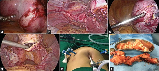
Figure 3.
(a) Intra-operative myomectomy being performed before hysterectomy in a case with large right lower uterine myoma extending into broad ligament. (b) 30° degree, 10 mm telescope camera skilfully rotated to visualize the left uterine artery from the left lateral aspect. (c) Myoma screw being used for manipulation and cranial traction during laparoscopic hysterectomy. (d) Vaginal vault opened posteriorly without any colpotomiser. This is then extended both sides to severe cervix from vault. (e) Port placement; 10 mm primary port at upper border of umbilicus, two 5 mm ports on the left side and one 5 mm port on the right side of abdomen. (f) Morcellated specimen depicting the technique to convert a globular specimen to longitudinal one

